There are four main categories of tissues found in the human body. They are Epithelial tissue, Connective tissue, Muscular tissue and Nervous tissue. Each one of these categories is defined by some unique characteristics.
For this lab you will need to follow the links on each of the histology pages to a virtual microscope that shows the various tissues. For each tissue there will be an unlabeled image, labeled image and video.
1) Start with the unlabeled image and zoom in move around and try and figure out what are some unique features.
2) After you feel you have an idea of a unique feature move to the labeled image and zoom in and move around. Notice the labeled features. These are the features that will help you identify the tissue. You do not need to know the terms of the labels. The labels are there to help show you the unique features of each tissue.
3) Optional. If you want the third link is a video of the tissue with a narration describing the tissue and helping explain identification of the tissue.
4) Complete the 'Student Report Form' after you have viewed and identified the histology images.
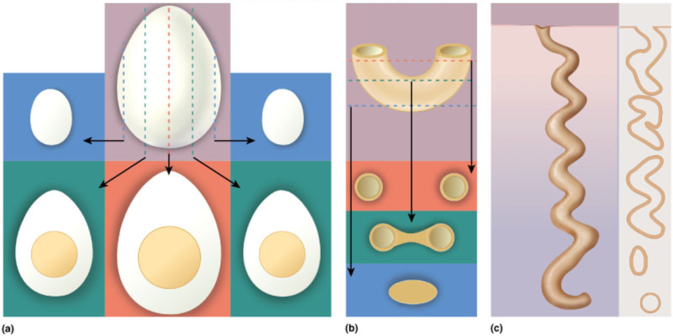
***As you look at the histology slides keep in mind that these are three dimensional tissues. That means that the particular section each slide is taken from may look a little different from another section of the same tissue. Take a look at the image below. You can see how the egg and noodle will look drastically different depending on where the exact cut is made. So if you see other histology images and they do not look exactly like what you see in this lab, that is perfectly normal. Every histology section will look slightly different. But the main features will always be there.
|