Move and zoom: drag the image to move it, or double-click to zoom in. Hold Ctrl (⌘ on a Mac) and scroll to zoom. On a touch screen, drag to move and pinch to zoom. With a keyboard, Tab to the image, then use the ← ↑ → ↓ keys to move, + and − to zoom, and 0 to reset. The buttons and slider above do the same thing, including the four arrow buttons for moving the image.
Open the full-size image in its own pageDescription of this slide
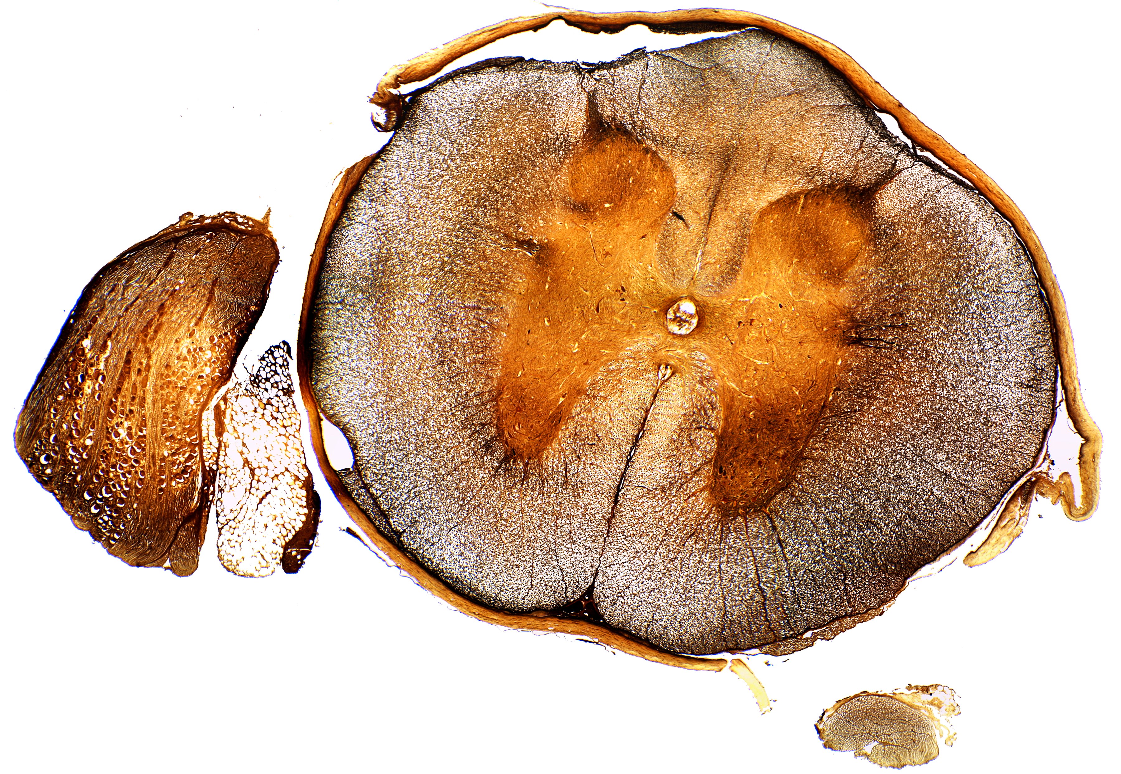
A cross-section of the spinal cord and some nearby tissue at low power, in orange-brown and gray tones, on a white background. A large, rounded, oval slice fills the center and right of the picture. Its outer ring is finely speckled gray-brown. A deep, narrow cleft cuts into the ring at the bottom center, and a shallow notch marks the top center. Inside the ring is a smoother, orange, butterfly-shaped area. It has two rounded lobes at the top and two thicker lobes at the bottom, joined across the middle. A tiny round hole sits at the center. A thin tan covering wraps around the outside of the slice and is pulled away at the upper left and the right. At the left of the slice are two separate pieces: an orange-brown oval piece full of round, pale cells, and beside it a small, pale piece full of white, bubble-like cells. A small, pale piece also lies at the lower right.