Move and zoom: drag the image to move it, or double-click to zoom in. Hold Ctrl (⌘ on a Mac) and scroll to zoom. On a touch screen, drag to move and pinch to zoom. With a keyboard, Tab to the image, then use the ← ↑ → ↓ keys to move, + and − to zoom, and 0 to reset. The buttons and slider above do the same thing, including the four arrow buttons for moving the image.
Open the full-size image in its own pageDescription of this slide
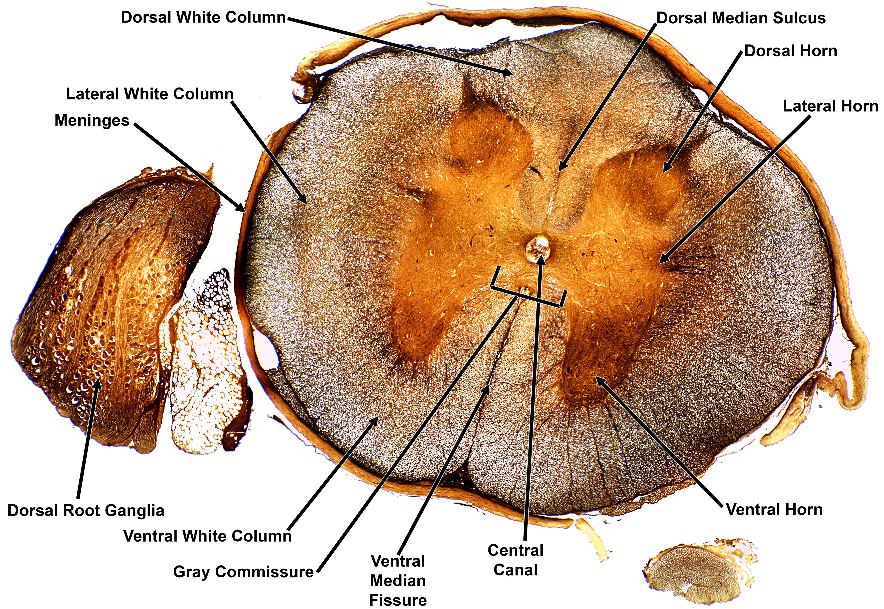
This is the same spinal cord slide as the unlabeled version, with twelve labels in black text, almost all with black arrows. The labels are Dorsal White Column, Dorsal Median Sulcus, Dorsal Horn, Lateral Horn, Lateral White Column, Meninges, Dorsal Root Ganglia, Ventral White Column, Gray Commissure, Ventral Median Fissure, Central Canal, and Ventral Horn.
Labels in this image
- Dorsal white column: the label is at the top left. One long arrow runs down and to the right, to the speckled gray-brown tissue at the top center of the slice, just left of the shallow notch.
- Dorsal median sulcus: the label is at the top right. One arrow runs down and to the left, to the narrow notch at the top center that runs down toward the middle of the slice.
- Dorsal horn: the label is at the top right, below the dorsal median sulcus label. One arrow runs down and to the left, to the rounded orange lobe at the upper right of the butterfly-shaped center.
- Lateral horn: the label is at the right, near the middle of the picture. One long arrow runs down and to the left, to the orange tissue at the right side of the butterfly-shaped center.
- Lateral white column: the label is at the upper left. One arrow runs down and to the right, into the speckled gray-brown outer ring on the left side of the slice.
- Meninges: the label is at the left. One arrow runs down and to the right, to the thin tan covering along the left edge of the slice.
- Dorsal root ganglia: the label is at the lower left. One arrow runs straight up, into the orange-brown piece to the left of the slice that is full of round, pale cells.
- Ventral white column: the label is at the bottom left. One arrow runs up and to the right, into the speckled gray-brown outer ring in the lower left part of the slice.
- Gray commissure: the label is at the bottom, left of center. One long arrow runs up and to the right, to the small notch just below the central hole. A small bracket also spans the narrow band of orange tissue just below that hole.
- Ventral median fissure: the label is at the bottom center, on three lines. One arrow runs up, to the deep, narrow cleft that cuts into the outer ring from the bottom center.
- Central canal: the label is at the bottom, right of center, on two lines. One arrow runs straight up, to the tiny round hole at the center of the butterfly-shaped area.
- Ventral horn: the label is at the lower right. One arrow runs up and to the left, to the thick orange lobe at the lower right of the butterfly-shaped center.