Move and zoom: drag the image to move it, or double-click to zoom in. Hold Ctrl (⌘ on a Mac) and scroll to zoom. On a touch screen, drag to move and pinch to zoom. With a keyboard, Tab to the image, then use the ← ↑ → ↓ keys to move, + and − to zoom, and 0 to reset. The buttons and slider above do the same thing, including the four arrow buttons for moving the image.
Open the full-size image in its own pageDescription of this slide
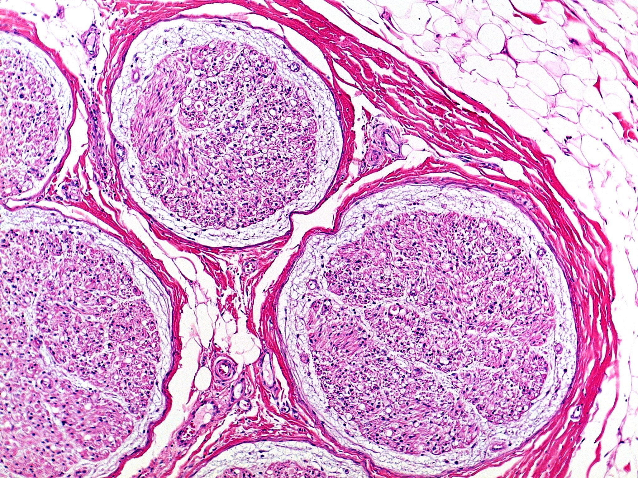
A nerve cut across at low power, stained pink and purple. Several large, round bundles fill the picture. Each is wrapped in a thin, pale layer and then a thick band of pink fibers. Inside each bundle is a fine, speckled mix of tiny round spaces, dots and small dark nuclei. One bundle is at the top center, a larger one at the lower right, another at the lower left, and parts of two more at the left edge and the bottom. Between the bundles are wavy pink fibers, pockets of white fat cells and small blood vessels. The upper right corner is filled with white, honeycomb-like fat cells, and a thick band of pink fibers curves along the right side.