Move and zoom: drag the image to move it, or double-click to zoom in. Hold Ctrl (⌘ on a Mac) and scroll to zoom. On a touch screen, drag to move and pinch to zoom. With a keyboard, Tab to the image, then use the ← ↑ → ↓ keys to move, + and − to zoom, and 0 to reset. The buttons and slider above do the same thing, including the four arrow buttons for moving the image.
Open the full-size image in its own pageDescription of this slide
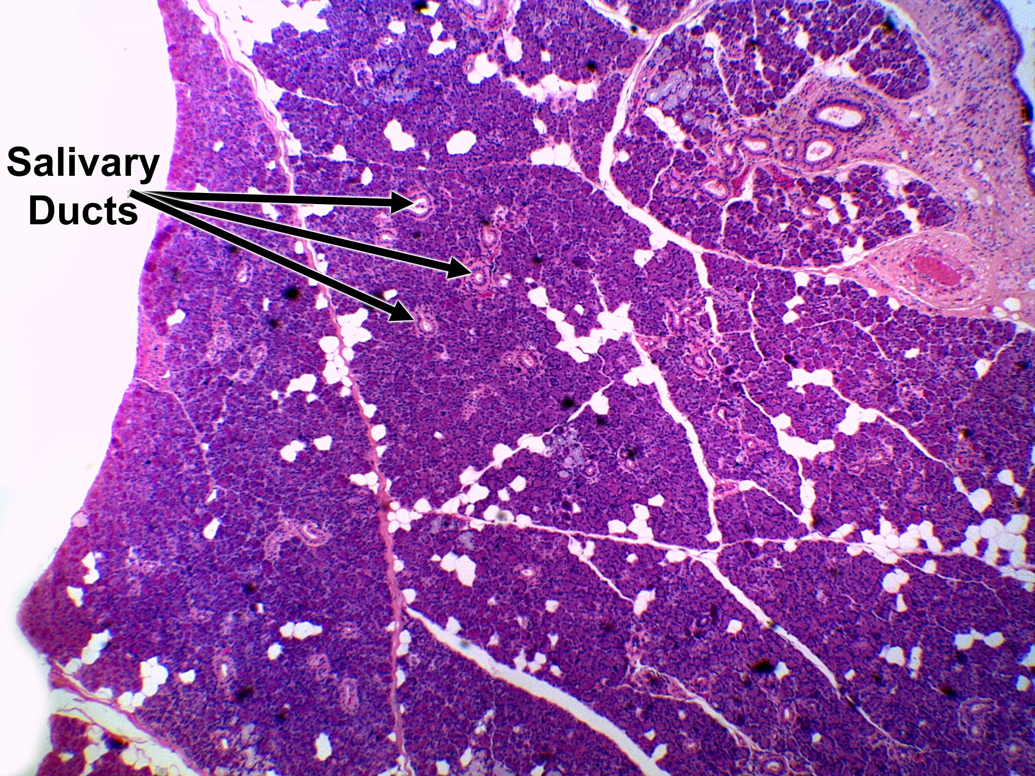
Low-power view of the parotid gland. Most of the tissue is densely packed, dark purple secretory tissue (serous acini only), divided into lobules by thin connective tissue. The salivary ducts are the small round or oval profiles with a clear central lumen and a pink wall. Larger ducts and a blood vessel lie in the connective tissue at the upper right. Scattered fat cells show as pale white spaces.