Move and zoom: drag the image to move it, or double-click to zoom in. Hold Ctrl (⌘ on a Mac) and scroll to zoom. On a touch screen, drag to move and pinch to zoom. With a keyboard, Tab to the image, then use the ← ↑ → ↓ keys to move, + and − to zoom, and 0 to reset. The buttons and slider above do the same thing, including the four arrow buttons for moving the image.
Open the full-size image in its own pageDescription of this slide
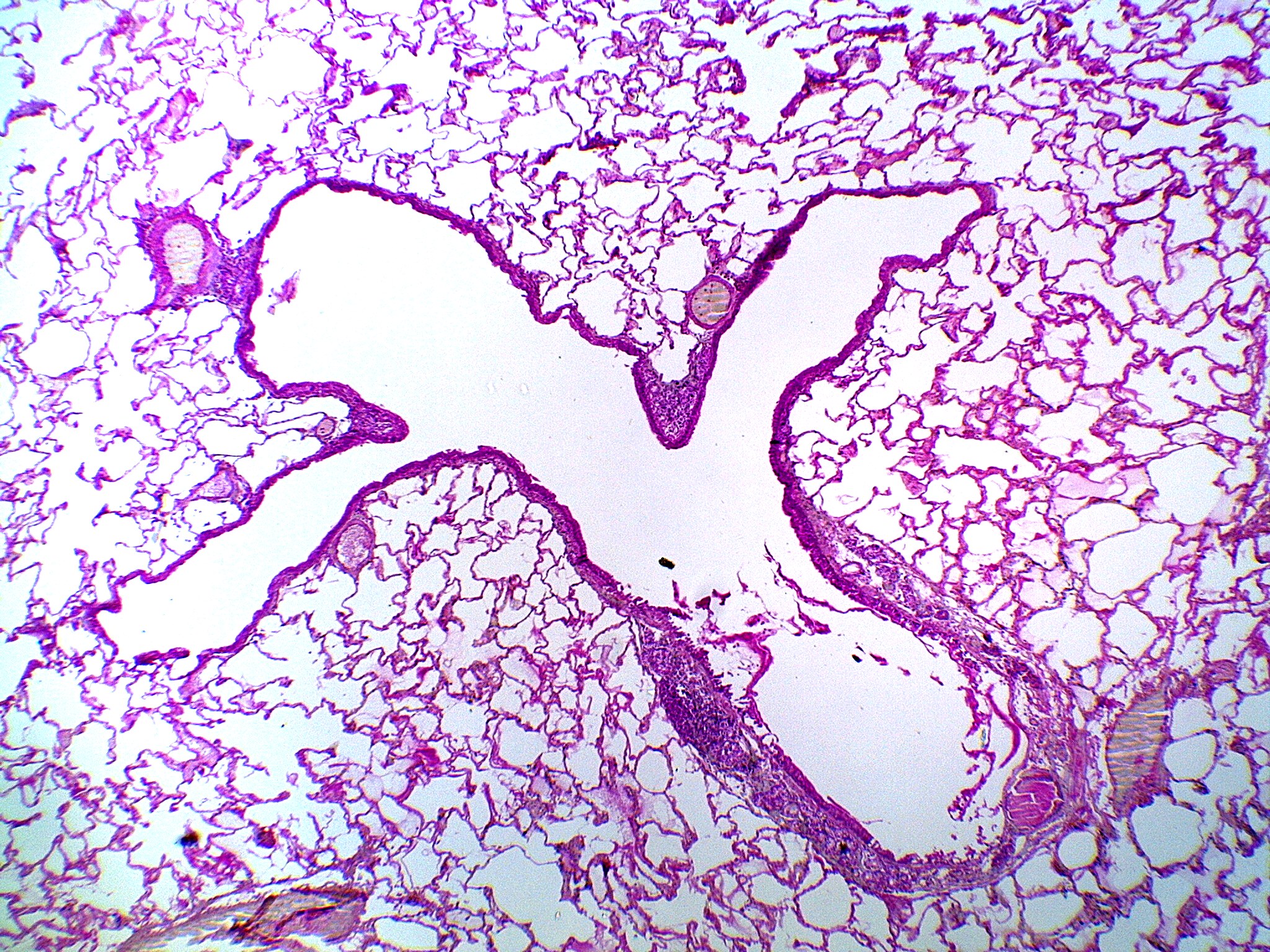
Low-power view of lung tissue showing the airway branching. The bronchiole (lower right) has a thicker wall and leads into the alveolar ducts, the wide, open passages that spread out through the center and left. The walls of an alveolar duct are mostly made of the openings of alveoli, so they look thin and broken up. Alveoli fill the rest of the field as a fine network of pink walls around empty air spaces.