Move and zoom: drag the image to move it, or double-click to zoom in. Hold Ctrl (⌘ on a Mac) and scroll to zoom. On a touch screen, drag to move and pinch to zoom. With a keyboard, Tab to the image, then use the ← ↑ → ↓ keys to move, + and − to zoom, and 0 to reset. The buttons and slider above do the same thing, including the four arrow buttons for moving the image.
Open the full-size image in its own pageDescription of this slide
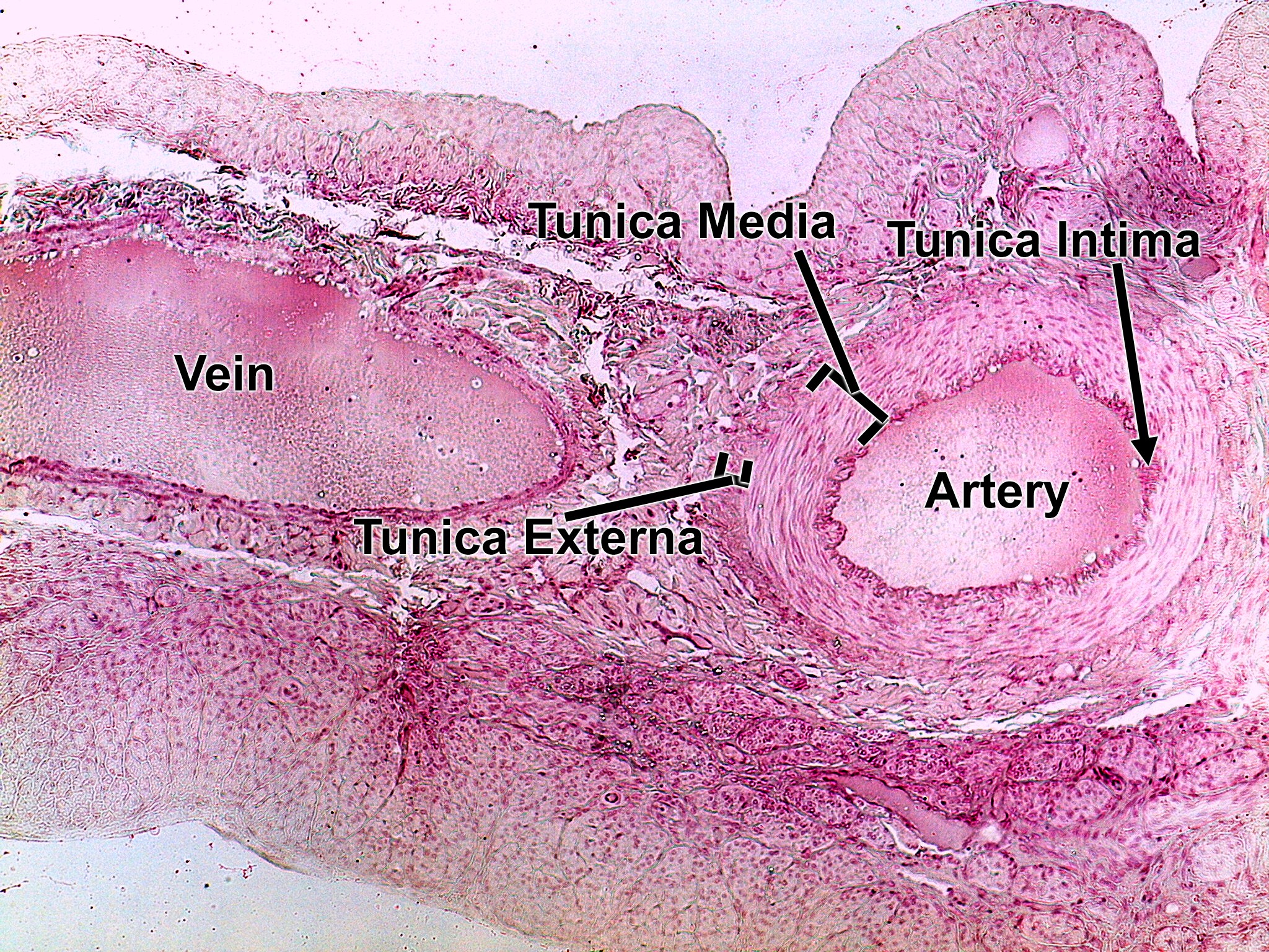
Low-power view of an artery and a vein in the same field, surrounded by other tissue. The artery (right) has a round lumen and a thick, muscular pink wall: the tunica intima lines the lumen, the thick tunica media is the middle layer, and the tunica externa blends into the surrounding connective tissue. The vein (left) has a very large, elongated lumen and a much thinner wall.