Move and zoom: drag the image to move it, or double-click to zoom in. Hold Ctrl (⌘ on a Mac) and scroll to zoom. On a touch screen, drag to move and pinch to zoom. With a keyboard, Tab to the image, then use the ← ↑ → ↓ keys to move, + and − to zoom, and 0 to reset. The buttons and slider above do the same thing, including the four arrow buttons for moving the image.
Open the full-size image in its own pageDescription of this slide
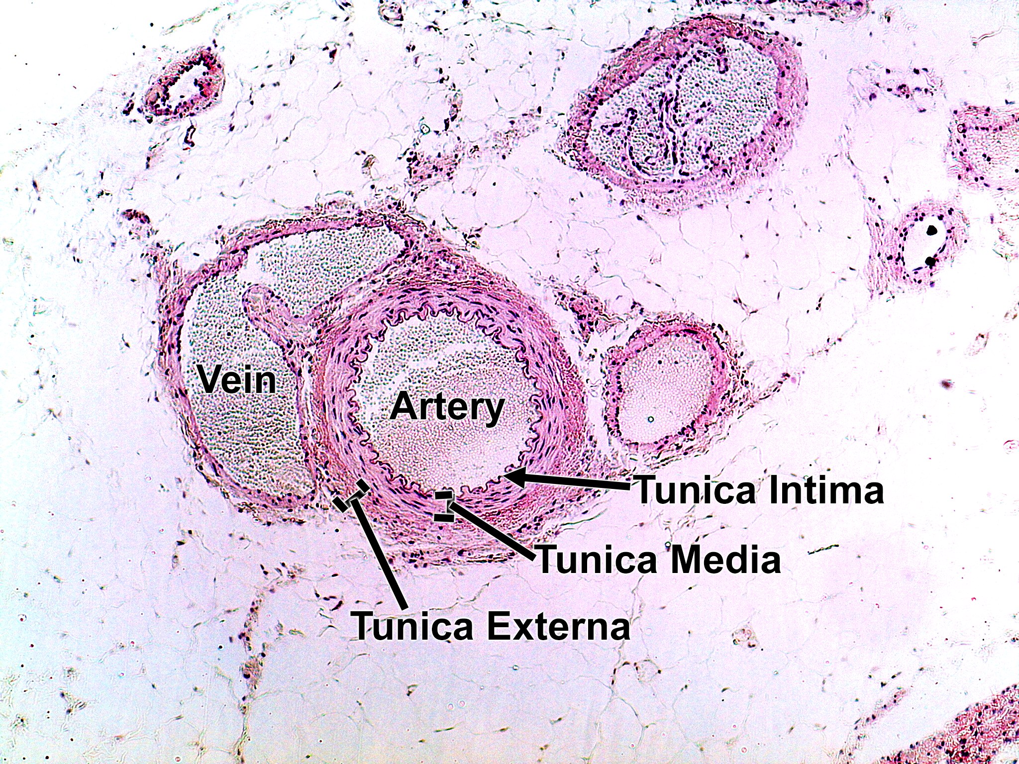
Low-power view of an artery and a vein lying side by side in fat tissue, with a few smaller vessels around them. The artery (center) has a round lumen and a thick wall with a wavy inner edge. From inside to outside the wall has the tunica intima, the tunica media (the thickest layer, made of smooth muscle) and the tunica externa. The vein (left) is next to it with a thinner wall and a larger, less regular lumen.