Move and zoom: drag the image to move it, or double-click to zoom in. Hold Ctrl (⌘ on a Mac) and scroll to zoom. On a touch screen, drag to move and pinch to zoom. With a keyboard, Tab to the image, then use the ← ↑ → ↓ keys to move, + and − to zoom, and 0 to reset. The buttons and slider above do the same thing, including the four arrow buttons for moving the image.
Open the full-size image in its own pageDescription of this slide
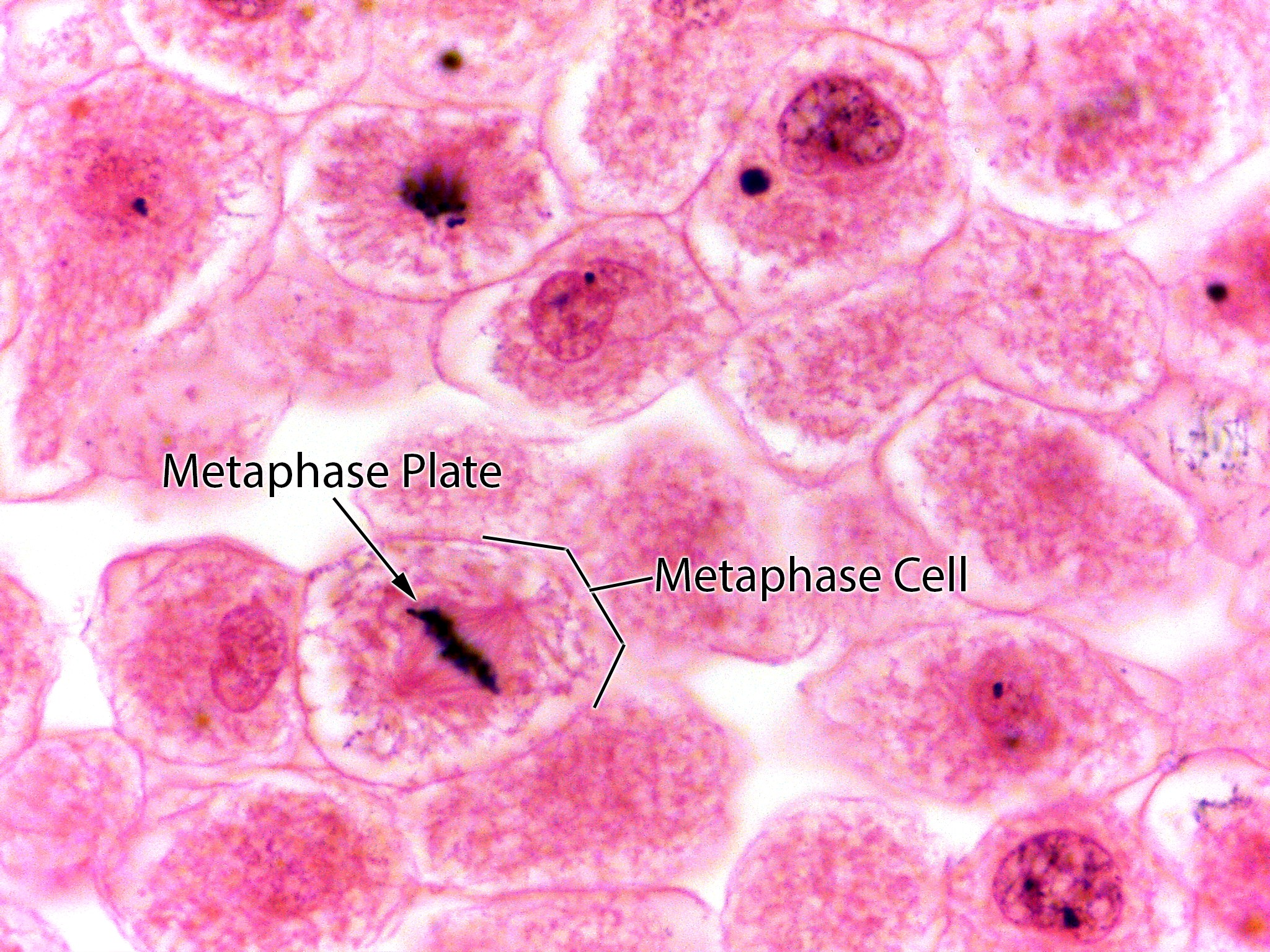
This is the same white fish slide as the unlabeled version, with two labels in black text. The labels are Metaphase Plate and Metaphase Cell.
Labels in this image
- Metaphase plate: the label is at the left of center, just above the cell at the lower left. An arrow points down and to the right from the label to the short, thick, dark band of chromosomes across the middle of that cell. This band is the metaphase plate.
- Metaphase cell: the label is at the right of center, in the middle of the picture. A short line leads from the label to a bracket along the upper right side of the cell at the lower left. The bracket marks that whole cell, the one with the dark band of chromosomes and fibers on both sides.