Move and zoom: drag the image to move it, or double-click to zoom in. Hold Ctrl (⌘ on a Mac) and scroll to zoom. On a touch screen, drag to move and pinch to zoom. With a keyboard, Tab to the image, then use the ← ↑ → ↓ keys to move, + and − to zoom, and 0 to reset. The buttons and slider above do the same thing, including the four arrow buttons for moving the image.
Open the full-size image in its own pageDescription of this slide
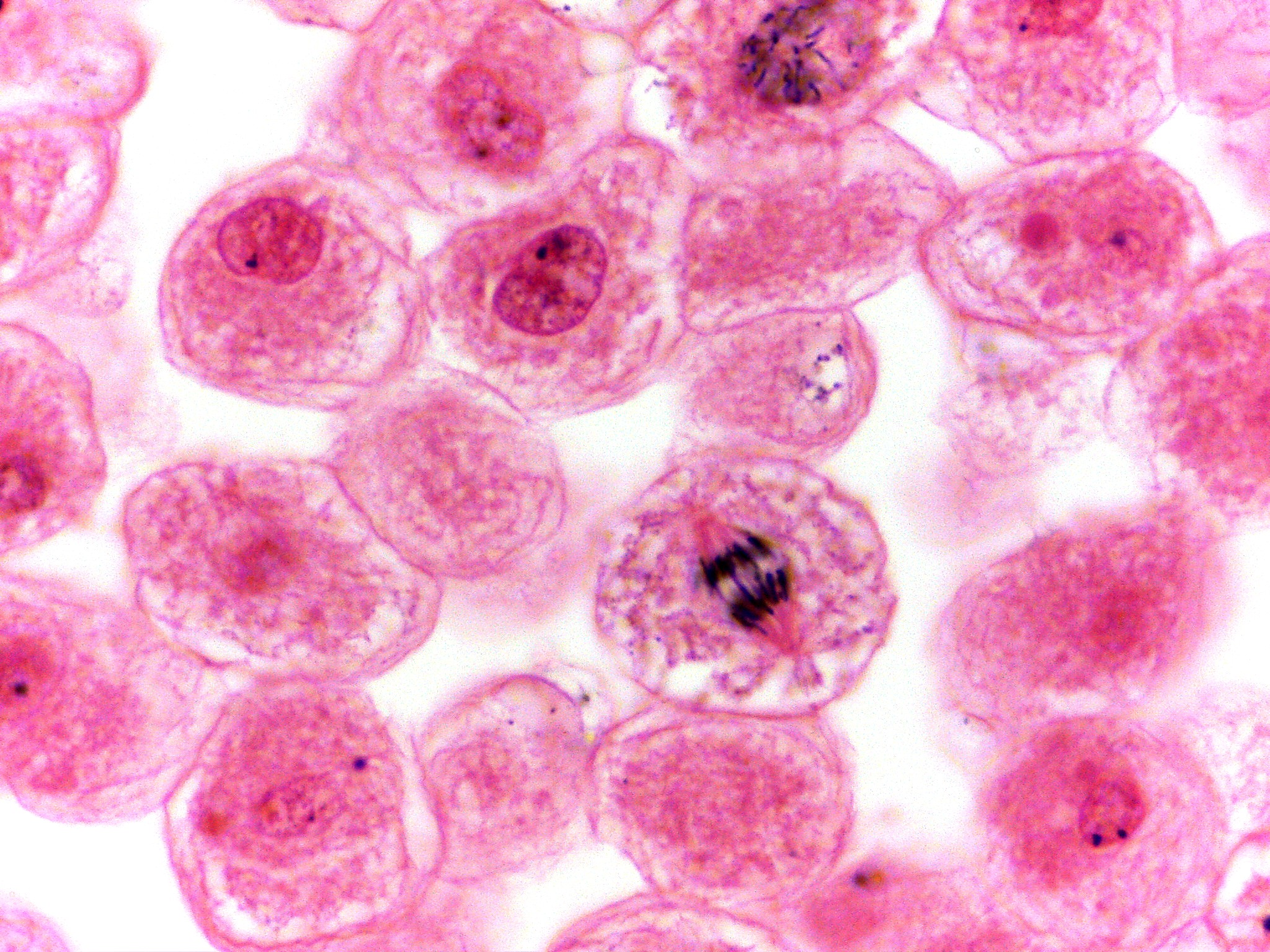
White fish cells at high power, stained pink. Large, round to oval, pale pink cells fill the picture, with white gaps between some of them. Several cells have a round, well-defined nucleus with a small dark dot inside, and others show only a faint, grainy center. One round cell near the lower center has no nucleus. Its cytoplasm looks finely speckled, and near its middle are two dark groups of rod-shaped chromosomes lying close together, just starting to separate. Pink fibers stretch from the chromosomes toward the upper left and lower right sides of the cell. A cell at the top of the picture also has a cluster of dark, thread-like chromosomes.