Move and zoom: drag the image to move it, or double-click to zoom in. Hold Ctrl (⌘ on a Mac) and scroll to zoom. On a touch screen, drag to move and pinch to zoom. With a keyboard, Tab to the image, then use the ← ↑ → ↓ keys to move, + and − to zoom, and 0 to reset. The buttons and slider above do the same thing, including the four arrow buttons for moving the image.
Open the full-size image in its own pageDescription of this slide
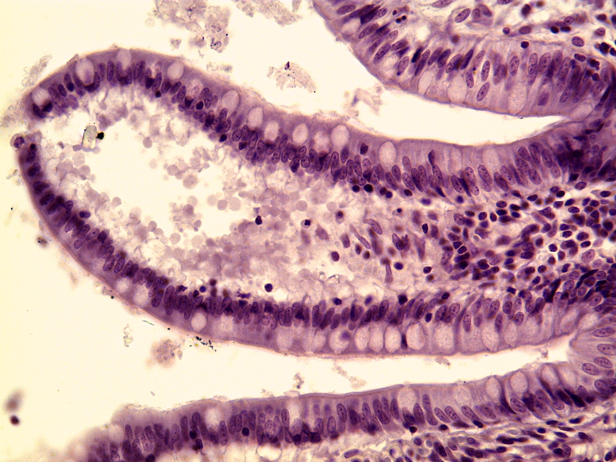
Lining of the small intestine (ileum) at high power, stained purple. A finger-shaped fold curves in from the left side of the picture. Its surface is covered by a single layer of tall, narrow cells standing side by side. Each tall cell has an oval, dark purple nucleus near its lower end, so the nuclei line up in a dark, wavy band along the edge of the tissue. Scattered among the tall cells are larger, rounded, pale cells with clear, almost empty centers. They show up along the top of the fold and along its lower edge. Inside the fold, small dark round nuclei of other cells crowd the center toward the right. Many pale, round, ghost-like shapes fill the open space at the left. Parts of neighboring folds appear at the top right and along the bottom.