Move and zoom: drag the image to move it, or double-click to zoom in. Hold Ctrl (⌘ on a Mac) and scroll to zoom. On a touch screen, drag to move and pinch to zoom. With a keyboard, Tab to the image, then use the ← ↑ → ↓ keys to move, + and − to zoom, and 0 to reset. The buttons and slider above do the same thing, including the four arrow buttons for moving the image.
Open the full-size image in its own pageDescription of this slide
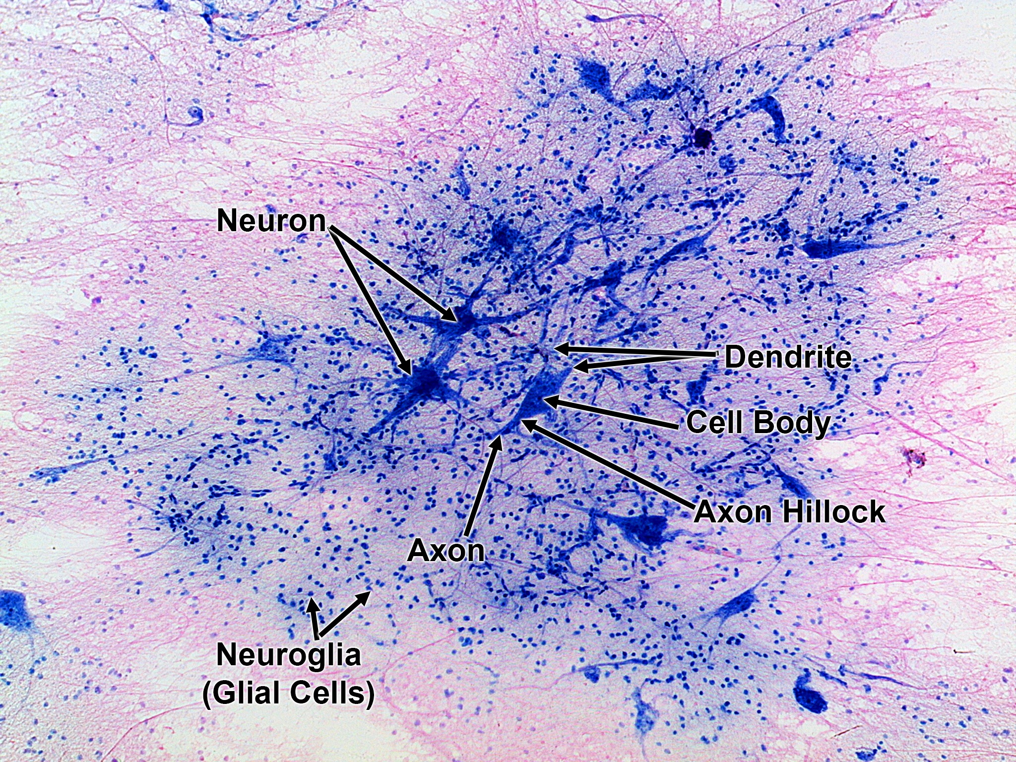
This is the same nervous tissue slide as the unlabeled version, with six labels in black text and arrows. The labels are Neuron, Dendrite, Cell Body, Axon Hillock, Axon and Neuroglia (Glial Cells).
Labels in this image
- Neuron: the label is at the upper left of center, in one line. Two arrows start together at its right end and spread apart, and both point down and to the right. One points to a star-shaped dark blue cell above and left of the center of the picture. The other points to a larger, darker star-shaped cell lower down, also left of center.
- Dendrite: the label is right of center, in one line. Two arrows start at its left end and run to the left, close together. They point to thin blue branches reaching out from the upper right of the dark cell in the middle of the picture.
- Cell body: the label is just below Dendrite, right of center, in two words. One arrow points to the left and slightly up, to the dark blue, rounded main part of the cell in the middle of the picture.
- Axon hillock: the label is lower right of center, in two words. One arrow points up and to the left, to the narrowing, cone-shaped part at the lower left of the same central cell, where it tapers into a thin fiber.
- Axon: the label is at the bottom center, in one word. One arrow starts above it and points up and slightly to the right, to the thin fiber that trails from the bottom of the central cell.
- Neuroglia (glial cells): the label is at the lower left, in two lines. Two short arrows start together just above it and spread apart. One points up, and the other points up and to the right. Both point to tiny blue dots, which are small cell nuclei, in the blue tissue near the lower left.