| 1 Timer |
| 1 Assistant |
| 1 Metric Ruler |
| Approximately 60cm length of vinyl tubing (basically enough to get from inside the contianer to the straw) |
| 2 Drinking straws |
| 1 - 3.8L (1 Gallon) clear plastic jug |
| 1 - 250mL measuring cup |
| 1 pail / contianer for water |
| Masking tape |
| Marking pen |
| Plastic tub (or can use sink if it can be stoppered) |
| Funnel |
| Food coloring (optional) |
| Dropper |
| Sponge |
Discussion and Review
Respiration is the exchange of gases between an organism and the external environment. The process of respiration is used in organisms to acquire oxygen that is used to provide energy.
Glucose, a simple sugar with the chemical equation C6H12O6, is made by plants through photosynthesis. Humans cannot produce their own energy, so they must obtain it from either plants or animals. Humans need oxygen to use the energy that comes from consuming other organisms. Some energy can be produced anaerobically – without oxygen – but the yield of energy is not as great as when using oxygen in aerobic respiration.
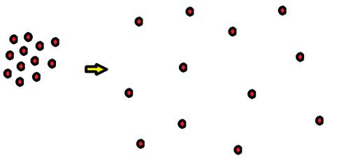
Humans undergo respiration in different ways, although all ways involve the intake of oxygen and release of carbon dioxide. This exchange of gas occurs because of diffusion. In diffusion, particles move from an area of high concentration to an area of low concentration. See Figure 1. For example, when a person inhales, the higher concentration of oxygen from the air enters the person's lungs, which contains less oxygen. Conversely, the higher levels of carbon dioxide in the lungs leaves and enters the air during exhalation.
Figure 1- Diffusion of Molecules from an area of high concentration (left) to an area of low concentration (right)
Humans undergo the following steps for respiration: ventilation, external respiration, and internal respiration.
- Ventilation – This is the cycle of inspired air and expired air. Inspired air is the air that goes from the external environment into the lungs. Expired air is the air that comes out of the lungs and moves to the external environment. Only organisms containing lungs experience ventilation.
- External respiration – This is the exchange of gases between the external environment, air or water, and the blood. In order for an organism to use external respiration, the epithelial tissue on organs such as lungs or gills – where gas is exchanged, must be:
- moist
- thin
- have a large surface area in relation to the organism itself
- Internal respiration – This is the exchange of gases within the body, for example, between cells and the blood.
Lungs - For humans, respiration includes ventilation, external respiration, and internal respiration. Air moves into the body through:
1. the nose and mouth,
2. the pharynx,
3. the larynx or vocal cords,
4. the trachea,
5. the right and left main bronchi
6. the bronchioles, and then finally to
7. the alveoli.
Air is warmed and humidified by the moist tissues as it moves through these passageways into the lungs. If any foreign particles enter the airway, they stick to the mucus in the trachea. Ciliated cells beat the mucus and particles upward toward the esophagus where they may be spit out or swallowed.
Figure 2 – Respiratory System, courtesy of Wikipedia Commons.
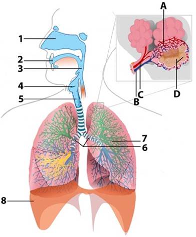
Table 2 – Items for Respiratory system
Item |
Description |
1 |
Nasal cavity |
2 |
Oral cavity |
3 |
Pharynx |
4 |
Larynx |
5 |
Trachea |
6 |
Main bronchi |
7 |
Bronchioles |
8 |
Diaphragm |
A |
Capillary beds |
B |
Pulmonary artery |
C |
Pulmonary vein |
D |
Alveoli |
|
|
|
|
|
|
|
|
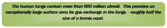 |
Once the air reaches the bronchi, it splits into smaller passages called bronchioles. Bronchioles end at sac-like structures called alveoli. Refer to Figure 5. The air that gets to the alveoli is 100% humidified, so the alveoli are moist. In addition, the membranes of the alveoli are very thin, and capillary beds that cover the alveoli are very thin. The oxygen and carbon dioxide gases must only cross two layers of cells – one layer of alveoli cells, and one layer of capillary cells – to exchange gases between the air and the blood. Because of this thin barrier, diffusion of the gases occurs very easily. The alveoli are circular in shape, so the surface area is increased. Since the concentration of oxygen is lower in the blood than in the air in the lungs, the oxygen readily moves into the blood. Conversely, since the carbon dioxide levels are much higher in the blood than in the air in the lungs, the carbon dioxide moves out of the blood into the air that will then be exhaled.
After the blood in the lungs gains oxygen and loses carbon dioxide, it flows to the heart where it then circulates to the rest of the body. The oxygen-rich blood flows through arteries, arterioles, and finally to the capillaries. The cells in the blood contain higher levels of oxygen than the cells of the body, so oxygen diffuses into the cells where it is needed. Since there is only one layer of epithelial cells lining the vessels, the oxygen easily diffuses out of the blood. The oxygen then enters the cells in the body. Carbon dioxide, which is a waste product in humans, diffuses out of the cells and into the blood. From the capillaries, the carbon dioxide–rich blood flows out into the capillaries, to venuoles and veins that carry blood back to the heart and pump it the lungs. See Figure 6. After in the lungs, the carbon dioxide may then be expired out of the body and into the air.
Figure 3 – Capillaries are small, thin vessels where gas exchange takes place easily.
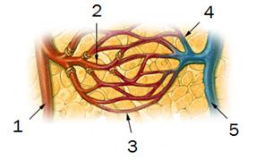
Table 3 – Items for Blood vessels
Item |
Description |
|
Item |
Description |
1 |
Arteries |
|
4 |
Venuoles |
2 |
Aterioles |
|
5 |
Veins |
3 |
Capillaries |
|
|
|
The amount of air respired may be an indicator of an individual's health. In the doctor's office, the device that measures the volume of air that moves in and out of the lungs is a spirometer. Complex methods of measuring full breaths and speed of breathing help determine if someone has diseased lungs.
However, if a spirometer is not available, as in the following exercise, the volume of air can also be measured – albeit less accurately – using a sinmple water volume setup. Measurement of the gases exchanged while at rest is called tidal volume. The amount of air respired can be calculated using simple means. This can be accomplished by measuring tidal volume measurements, and recording the average breaths taken per minute. The amount of air respired per minute can also be called minute ventilation (VE).
The measure of the maximum volume of air breathed in and out is called forced vital capacity. From the values obtained through the forced expiratory volume measurements, a doctor can predict the volume of air that the lungs can hold. NOTE: There is always some air that remains in the lungs, called the residual volume. This value must be added in after finding the forced vital capacity, as will be done in the following exercise.