Discussion and Review
Mitosis is the process by which the body builds tissue and by which some organisms reproduce asexually. All biological eukaryotic organisms undergo mitosis for growth and repair. The purpose of mitosis is to create an identical replica of a cell. To undergo mitosis, the chromosomes and organelles must be copied. This duplication will ensure that when the cell divides, each new cell has the same genetic information and contents as the parent cell. The result yields two genetically identical daughter cells. In some cells, such as prokaryotic cells, the division of cells is called fission.
The terms haploid and diploid refer to the number of chromosome sets in a cell. Diploid (abbreviated as 2n) organisms have two sets of chromosomes. Human cells (except for gametes, discussed below), most animal cells, and many plant cells are diploid. Haploid (abbreviated as n) cells such as eggs or sperm have only one set of chromosomes. Refer to Figure 1. Homologous chromosomes are sets of chromosomes that have the same genes (normally one chromosome from the mother, one from the father).
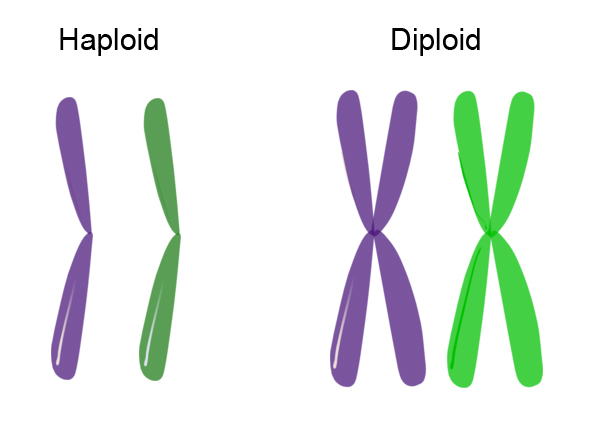
Figure 1. Haploid versus diploid.
Sex cells divide through a different form of cell division called meiosis in which the final cells have only half the number of chromosomes as the parent cell. Meiosis is the process by which sperm and eggs are produced and involves two separate nuclear divisions. The resultant cell is haploid (n).Table 1 compares and contrasts characteristics of mitosis and meiosis.
| |
Mitosis |
Meiosis |
Chromosome number of parent cells |
2n |
2n |
Number of DNA replications |
1 |
1 |
Number of divisions |
1 |
2 |
Number of daughter cells produced |
2 |
4 |
Chromosome number of daughter cells |
2n |
n |
Purpose |
Growth and repair |
Gamete and spore production |
Table 1. Mitosis versus meiosis.
During mitotic cell division, cells replicate themselves while maintaining the same number of homologous chromosome pairs, which have the same characteristics and the same lengths. Mitosis involves the duplication of a cell's chromosomes via the unzipping of the chromosomes of the original parent cell and the orderly re-zipping of the old chromosomes and newly replicated chromosomes into two exact copies of the original cell.
Mitosis is one part of the continuously reoccurringcell cycle responsible for growing, replacing, and renewing cells in eukaryotes. The normal cell cycle is comprised of two main stages: mitosis (cell division), and interphase (the time between cell divisions). See Figure 2. In preparation for mitosis, the cell duplicates the DNA and most cell organelles.
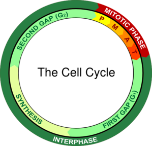
Figure 2. Diagram of the cell cycle.
Stages of Mitosis
The process of mitosis is continuous, and it happens at different stages in specific parts of plants or animals. The process of mitosis is complex, and has four distinct phases:
- In prophase, chromosomes are condensed in the nucleus and become visible under a microscope. The envelope around the nucleus disintegrates, the nucleolus disappears, and mitotic spindles begin forming at opposite poles of the cell. Mitotic spindles are composed of many fibers, including microtubules. Microtubules are components of the cell's cytoskeleton. The cytoskeleton is a dynamic component of the cell: it maintains cellular shape and enables various forms of movement and transport. Centromeres and centrioles organize the mitotic spindles. See Figure 3.
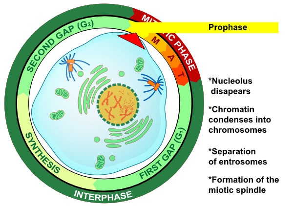
Figure 3. Prophase: the first stage of mitosis.
- Metaphase begins as the replicated chromosomes move toward the center of the cell. This movement is coordinated by the microtubule-based spindle fibers which are anchored to the centromeres and centrioles. Metaphase ends with the chromosomes arranging themselves along the equatorial plane of the cell. See Figure 4.
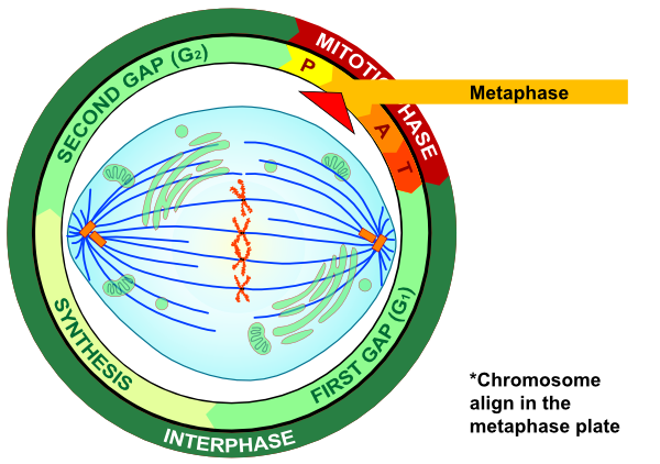
Figure 4. Metaphase: the second stage of mitosis.
- During anaphase, the spindle fibers begin to shorten and pull the sister chromatids— identical portions of the chromosomes—apart. Subsequently, the centromeres break and the replicated chromatids separate, creating two separate and identical chromosomes. As anaphase progresses, the chromosomes are pulled further apart. See Figure 5.
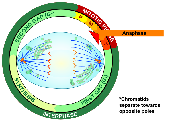
Figure 5. Anaphase: the third stage of mitosis.
- Telophase is the last stage of mitosis. The two sets of separated chromosomes elongate and are grouped into new nuclei surrounded by a nuclear envelope. At the close of telophase, the mitotic process of replicating, dividing, and then re-pairing the chromosomes is immediately followed by cytokinesis. During cytokinesis, the cell divides the cytoplasm, which is the watery substance that contains other cellular components important to the functioning of the cell. A new cell wall and/or membrane is produced which divides the two new cells. In animals (see Figure 6), this process is accomplished through the formation of a cleavage furrow, followed by the pinching off of new cells. See Figure 7.
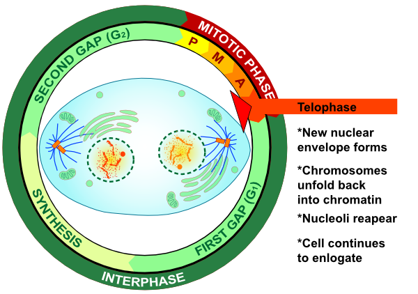
Figure 6. Telophase: the fourth and final stage of mitosis.
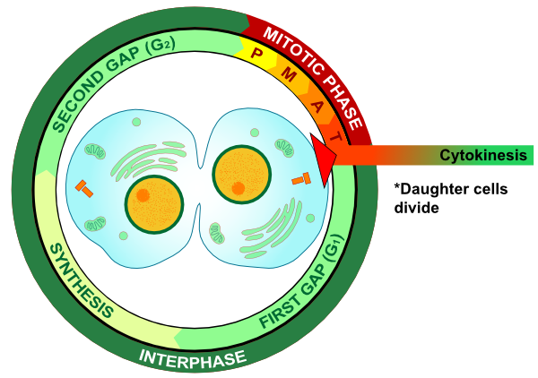
Figure 7. Cytokinesis -- Causes the actual separation of the cell membranes.
***Look through the various stages of mitosis as seen through a microscope of onion root tips and white fish cells here.***
While mitosis takes place throughout an organism's body, mitotic cell division rates increase at injury sites to assist in the repair and replacement of damaged cells. Some areas in the body show higher mitosis rates than others. For example, skin cells have a high rate of mitosis and chondrocytes (cartilage cells) have a low rate of mitosis. Mitotic cell division also occurs at a higher rate in infants and young children, as they grow rapidly and show an increased need for new cells.
Cell division rates vary: Some cells such as neurons are not able to divide at all, while epithethial tissue cells lining the stomach have very high division rates. It is possible to quantify the rate of a dividing cell population and then examine and evaluate the differences in various cells' capabilities to divide. This quantification allows the dynamics of cell division to be evaluated in different and/or adjacent cell areas of an organism.
The rate of mitosis changes in an organism depending on the location and function of the cells within that organism. If growth is needed more rapidly in one area, such as in the root of a plant, the percentage of cells within that area will increase in mitotic activity. The mitotic index is the calculation of this rate, using the following equation:
Mitotic Index = (Number of cells in mitosis / Total number of cells) × 100
In this experiment, you will observe pictures taken from the end of a plant root, which is also called the apical meristem. Under the apical meristem is the root cap, which protects the rapidly growing area. Gravity guides the growth of the root.
Figure 8 was derived from mitotic index measurements taken from cell areas at various distances from the root cap. It shows that the mitotic index decreases as the distance from the root cap increases and that mitotic activity increases toward root tips. Mitotic index comparisons are often used to evaluate the impact of environmental factors on plants. For example, plants grown on a vehicle in spaceflight have a higher mitotic index than control plants grown on the ground. Mitotic index measurements are also very valuable in medical research and are a key factor in evaluating the growth of various cancers.
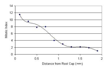
Figure 8. Mitotic index measurements at distances from the root cap of a plant.
|