Move and zoom: drag the image to move it, or double-click to zoom in. Hold Ctrl (⌘ on a Mac) and scroll to zoom. On a touch screen, drag to move and pinch to zoom. With a keyboard, Tab to the image, then use the ← ↑ → ↓ keys to move, + and − to zoom, and 0 to reset. The buttons and slider above do the same thing, including the four arrow buttons for moving the image.
Open the full-size image in its own pageDescription of this slide
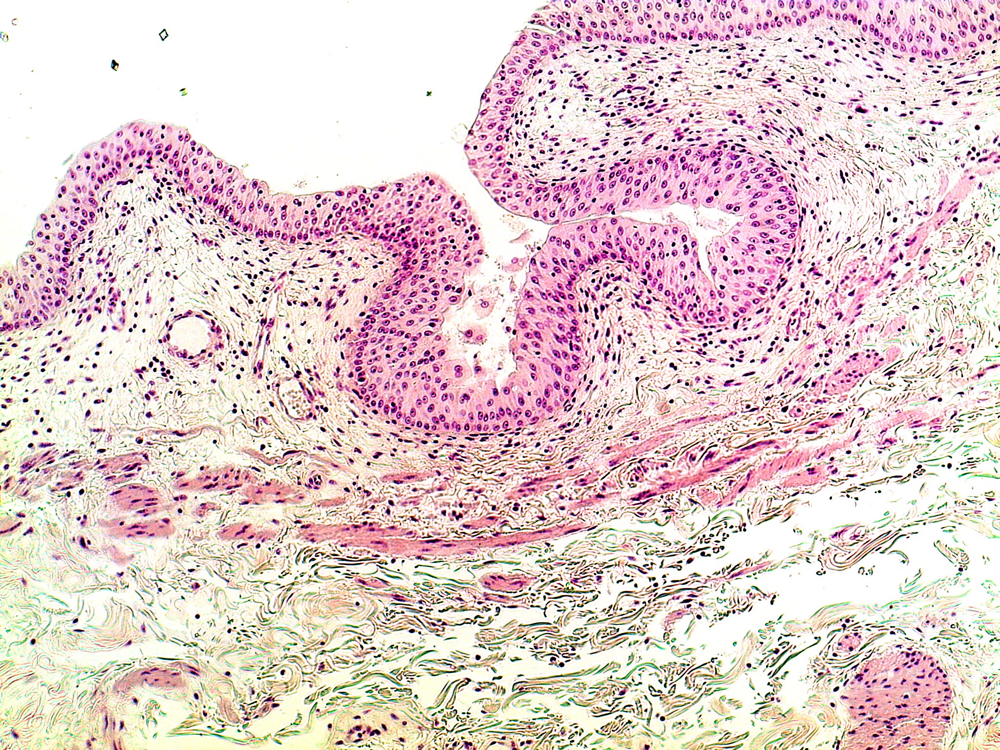
Medium-power view of the bladder wall. The lining is transitional epithelium: a thick, many-layered pink epithelium with round nuclei, with larger rounded cells at the surface. It is folded because the bladder is empty. Below it is loose connective tissue with small vessels, then bundles of smooth muscle.