Move and zoom: drag the image to move it, or double-click to zoom in. Hold Ctrl (⌘ on a Mac) and scroll to zoom. On a touch screen, drag to move and pinch to zoom. With a keyboard, Tab to the image, then use the ← ↑ → ↓ keys to move, + and − to zoom, and 0 to reset. The buttons and slider above do the same thing, including the four arrow buttons for moving the image.
Open the full-size image in its own pageDescription of this slide
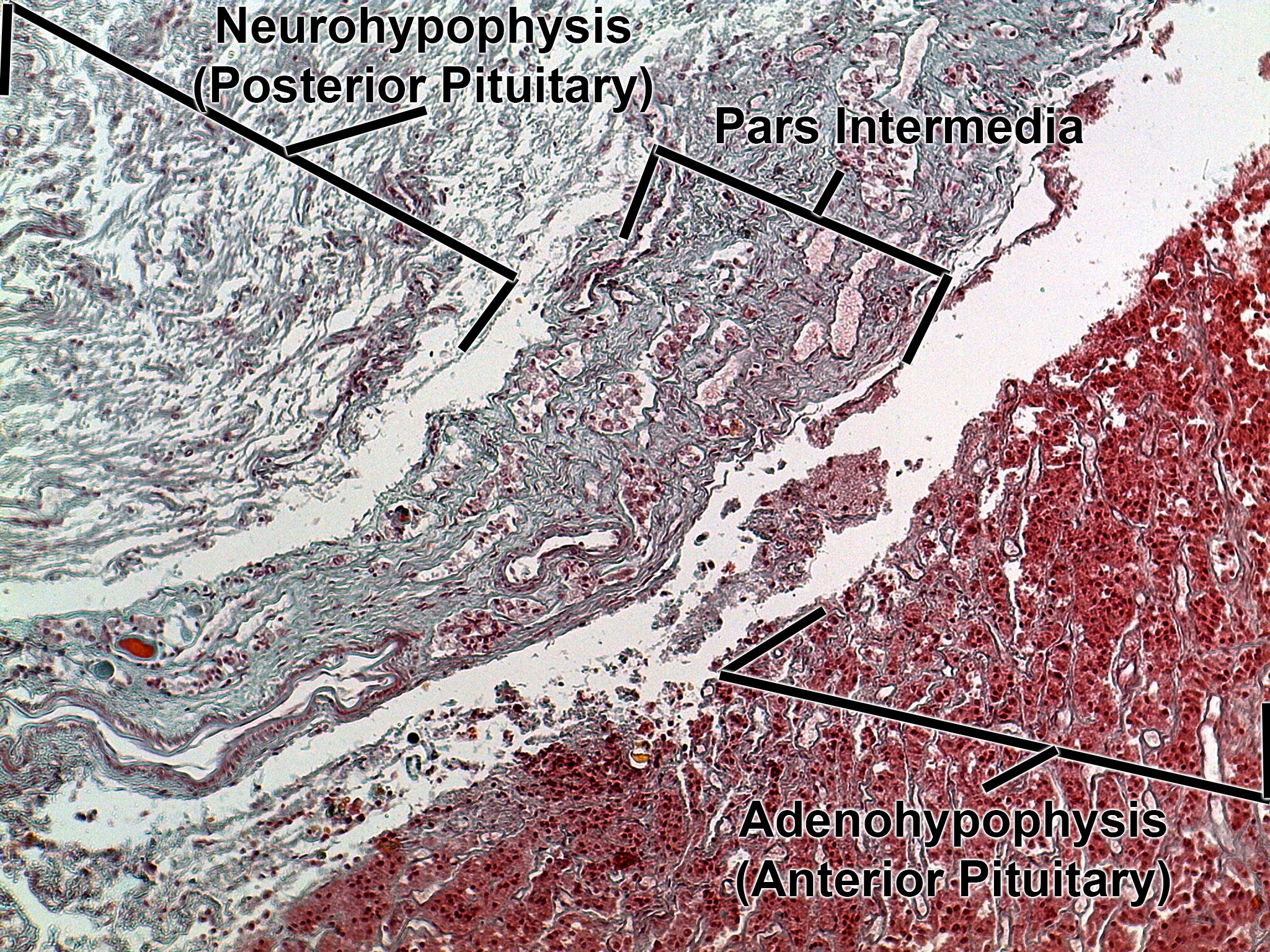
Low-power view with a different stain (red and blue-green). The adenohypophysis (anterior pituitary) is the dense, red-stained cellular tissue. The pars intermedia is the narrow band of blue-green connective tissue with small cell clusters and follicle-like spaces, next to the pale gap. The neurohypophysis (posterior pituitary) is the loose, pale, fibrous tissue.