Move and zoom: drag the image to move it, or double-click to zoom in. Hold Ctrl (⌘ on a Mac) and scroll to zoom. On a touch screen, drag to move and pinch to zoom. With a keyboard, Tab to the image, then use the ← ↑ → ↓ keys to move, + and − to zoom, and 0 to reset. The buttons and slider above do the same thing, including the four arrow buttons for moving the image.
Open the full-size image in its own pageDescription of this slide
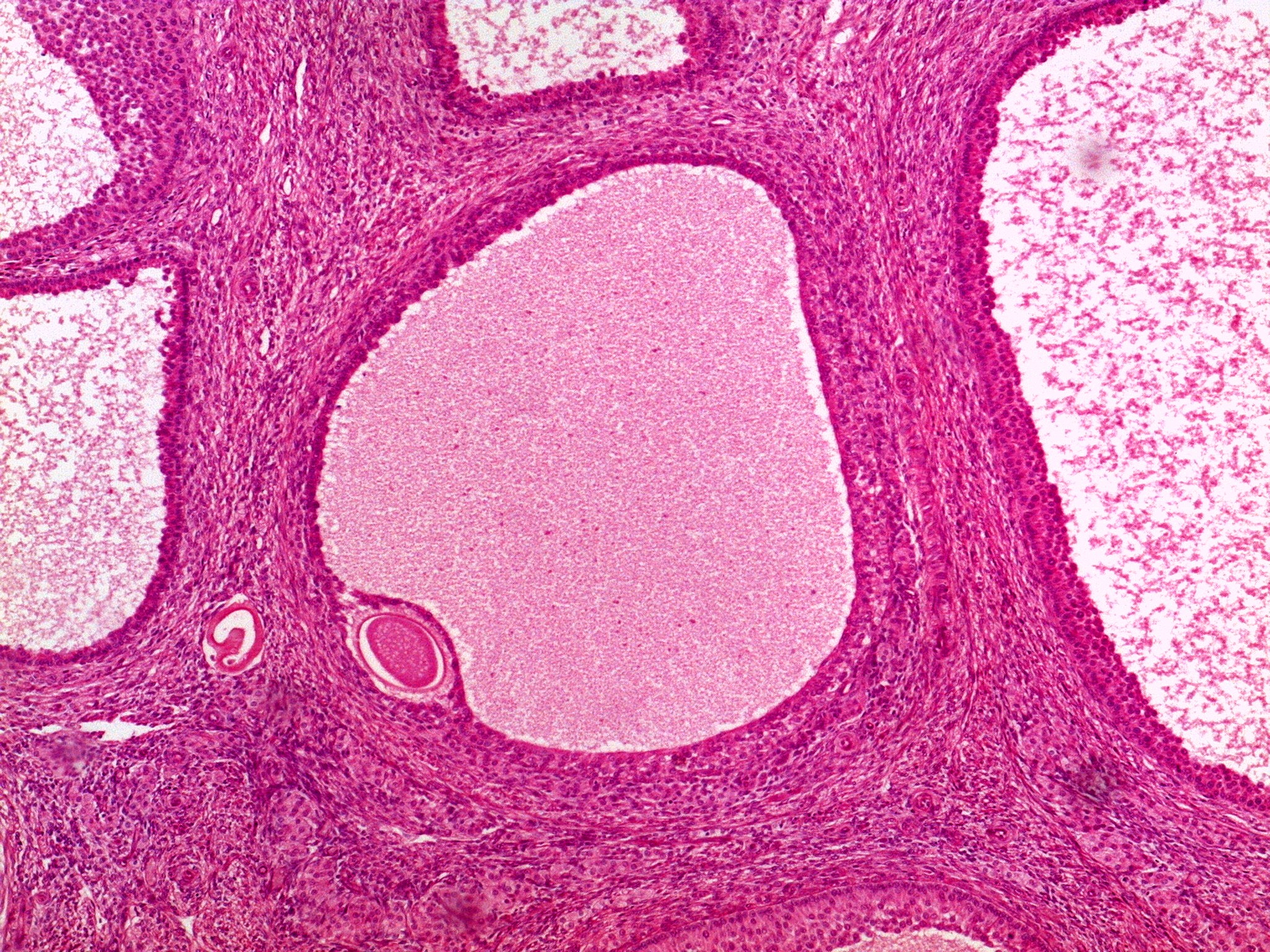
Low-power view of a vesicular (Graafian) follicle. Most of the follicle is the huge, pale pink antrum filled with fluid. The wall around it is made of follicular (granulosa) cells with connective tissue layers outside. The oocyte, surrounded by the zona pellucida, sits at the lower left edge of the antrum.