Move and zoom: drag the image to move it, or double-click to zoom in. Hold Ctrl (⌘ on a Mac) and scroll to zoom. On a touch screen, drag to move and pinch to zoom. With a keyboard, Tab to the image, then use the ← ↑ → ↓ keys to move, + and − to zoom, and 0 to reset. The buttons and slider above do the same thing, including the four arrow buttons for moving the image.
Open the full-size image in its own pageDescription of this slide
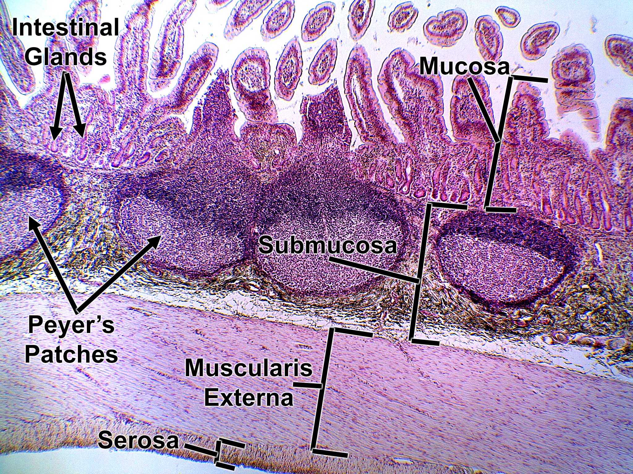
Low-power view of the ileal wall. The mucosa along the top is made of many villi, with the short intestinal glands at their bases. The Peyer’s patches are the large, round, dark purple clusters of lymphoid tissue (lymphatic nodules), the key feature of the ileum. They sit in the mucosa and push into the pale submucosa. Below them are the thick, pink muscularis externa and the thin serosa.