Move and zoom: drag the image to move it, or double-click to zoom in. Hold Ctrl (⌘ on a Mac) and scroll to zoom. On a touch screen, drag to move and pinch to zoom. With a keyboard, Tab to the image, then use the ← ↑ → ↓ keys to move, + and − to zoom, and 0 to reset. The buttons and slider above do the same thing, including the four arrow buttons for moving the image.
Open the full-size image in its own pageDescription of this slide
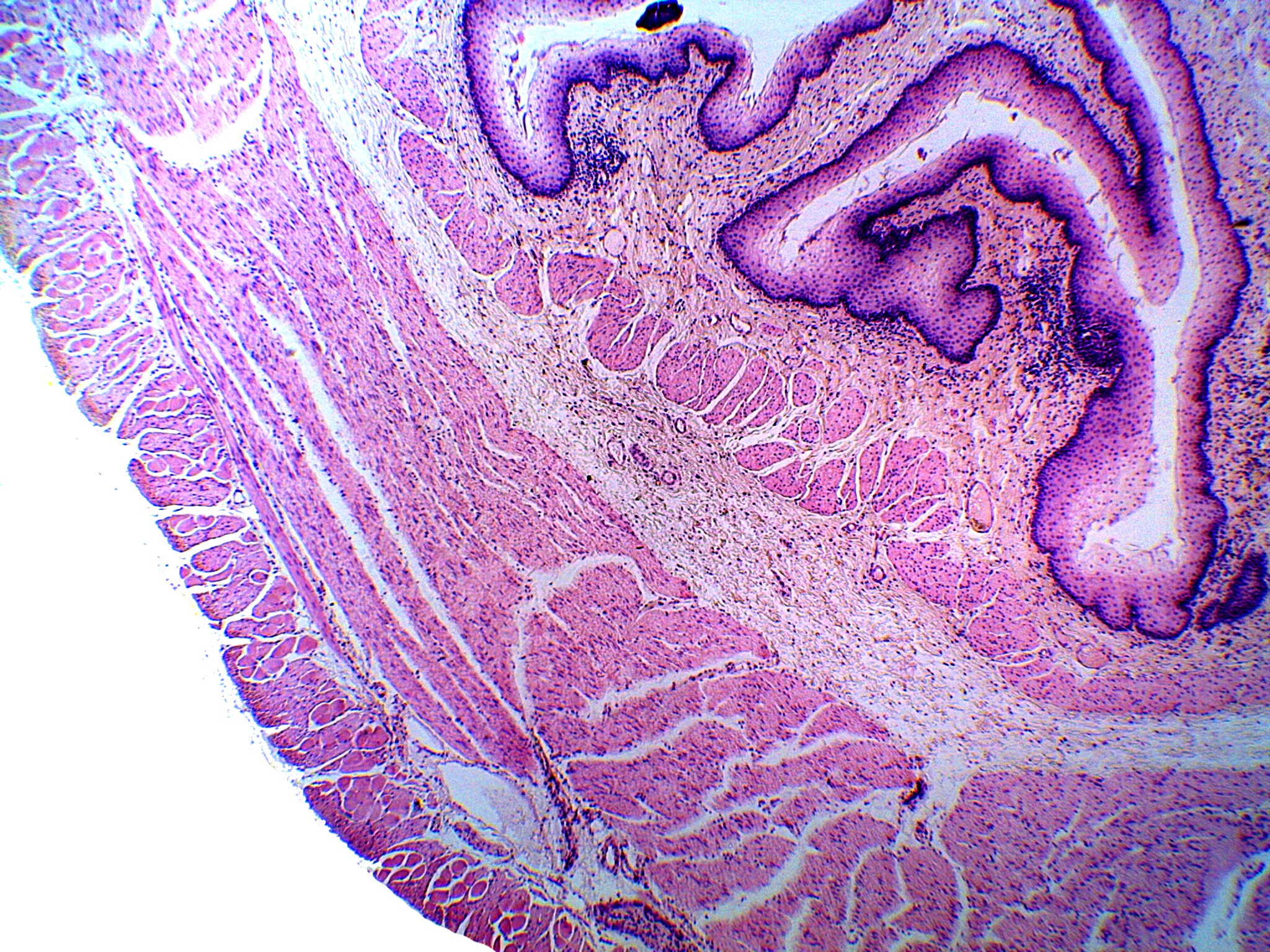
Low-power view of the esophageal wall cut across, with the lumen toward the upper right. From the lumen outward: the mucosa has a thick, dark-staining stratified squamous epithelium, a pale lamina propria, and a thin muscularis mucosae. Next is the pale submucosa. The muscularis externa has an inner circular layer and an outer longitudinal layer. The outermost layer is the adventitia. The mucosa is folded, so the lumen is irregular.