Move and zoom: drag the image to move it, or double-click to zoom in. Hold Ctrl (⌘ on a Mac) and scroll to zoom. On a touch screen, drag to move and pinch to zoom. With a keyboard, Tab to the image, then use the ← ↑ → ↓ keys to move, + and − to zoom, and 0 to reset. The buttons and slider above do the same thing, including the four arrow buttons for moving the image.
Open the full-size image in its own pageDescription of this slide
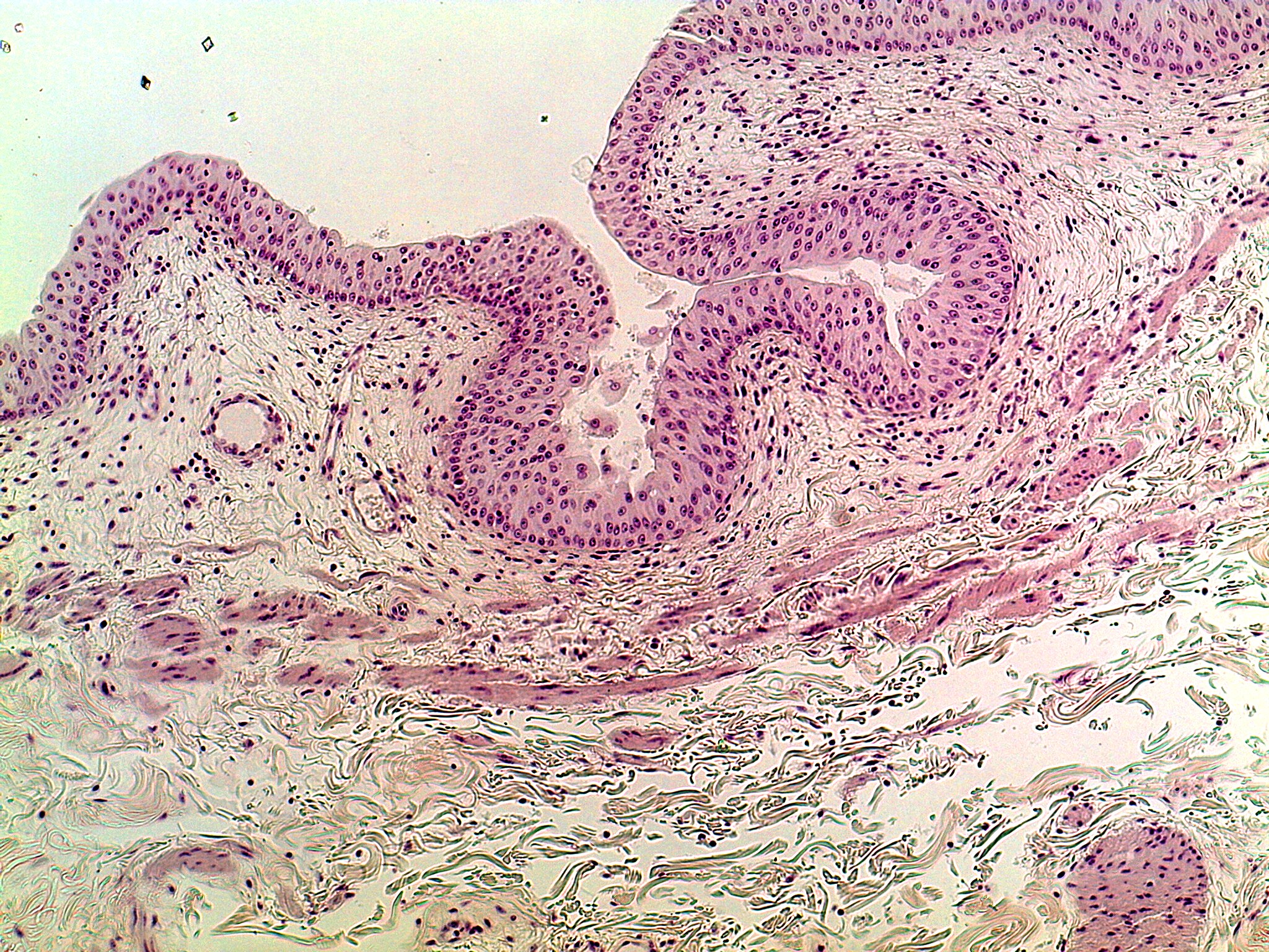
Wall of the urinary bladder at low power, stained pink and purple, with a pale cream background at the upper left. The lining forms wavy folds. It is several cells thick, and its round to oval nuclei are scattered at different levels. A ridge at the upper left curves down toward the center, a large folded loop at the center wraps around an open space that holds pale debris, and a second fold fills the upper right. Below the lining is pale, loose tissue with scattered nuclei and a small round blood vessel at the left. Bands of pink-purple, stretched-out muscle run diagonally across the lower middle. Wispy fibers fill the bottom of the picture, with another piece of lining in the lower right corner.