Move and zoom: drag the image to move it, or double-click to zoom in. Hold Ctrl (⌘ on a Mac) and scroll to zoom. On a touch screen, drag to move and pinch to zoom. With a keyboard, Tab to the image, then use the ← ↑ → ↓ keys to move, + and − to zoom, and 0 to reset. The buttons and slider above do the same thing, including the four arrow buttons for moving the image.
Open the full-size image in its own pageDescription of this slide
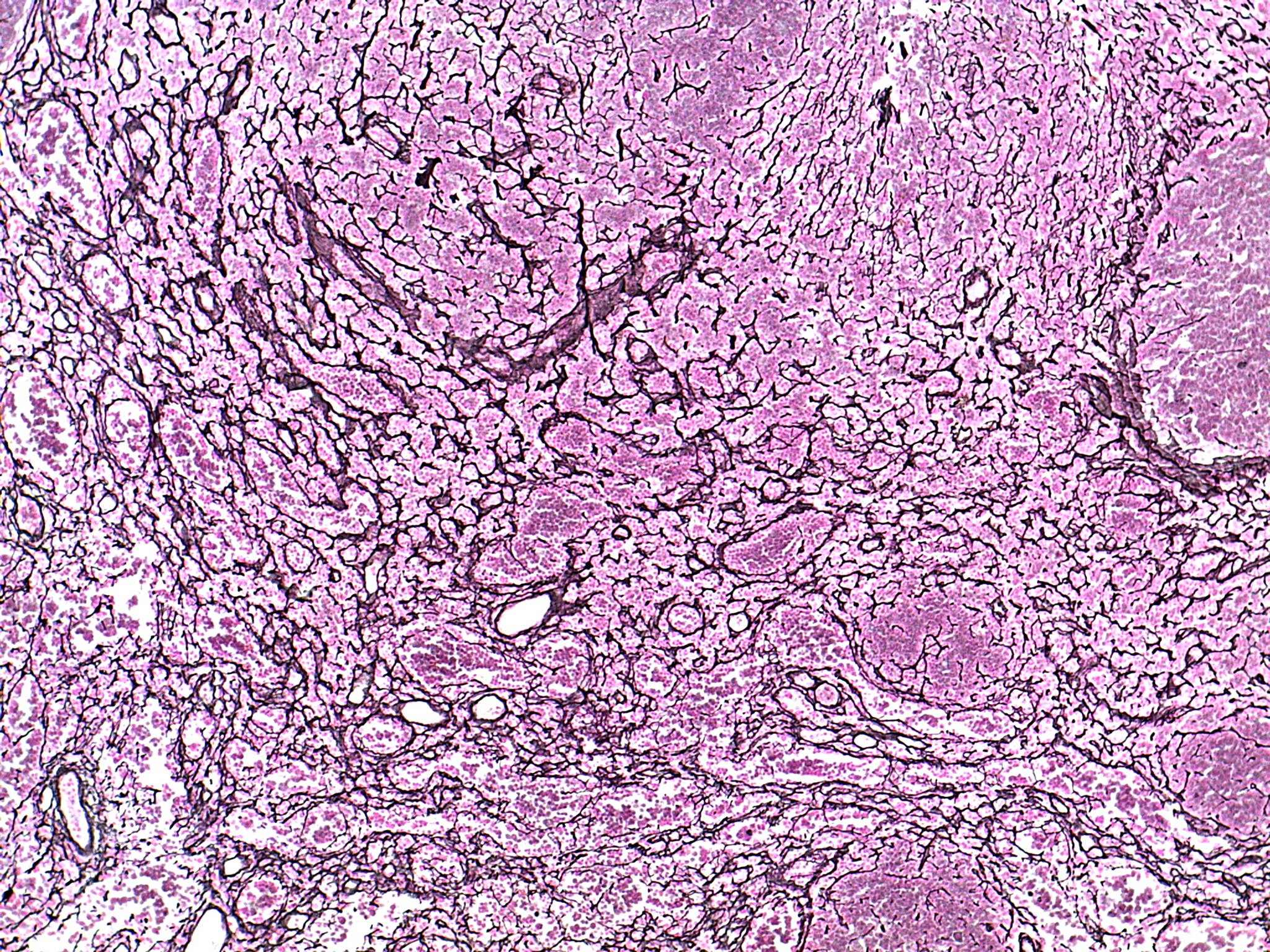
Spleen tissue at low power, stained pink and purple with a special stain that makes the fine fibers black. A net of thin black fibers covers the whole picture. The spaces in the net are filled with pink, grainy clusters of cells. The net is dense and crowded along the left side and the lower half, and looser in the upper middle. A few round, clear white openings sit near the lower center and the lower left corner, and a large, darker purple, rounded area fills the right edge.