Move and zoom: drag the image to move it, or double-click to zoom in. Hold Ctrl (⌘ on a Mac) and scroll to zoom. On a touch screen, drag to move and pinch to zoom. With a keyboard, Tab to the image, then use the ← ↑ → ↓ keys to move, + and − to zoom, and 0 to reset. The buttons and slider above do the same thing, including the four arrow buttons for moving the image.
Open the full-size image in its own pageDescription of this slide
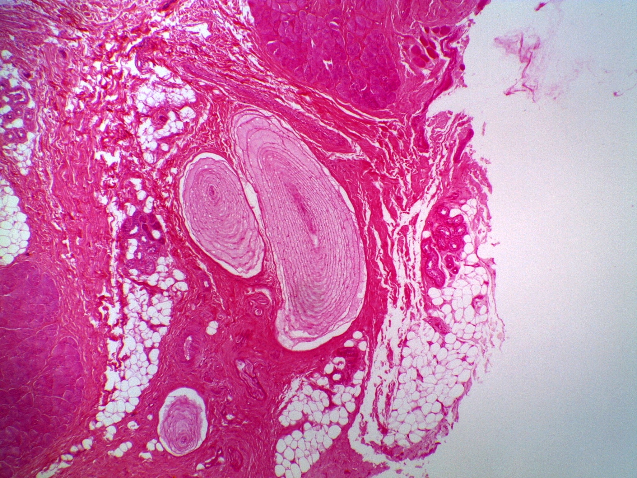
Skin tissue from just under the surface, at low power, stained deep pink and red. The right quarter of the picture is pale, with one torn pink wisp near the top right corner. At the center are two large, egg-shaped structures built of many thin, pale pink, circular layers, like the layers of an onion. The larger one is long and tilted. The smaller, rounder one sits just to its left. A thin white rim edges each of them. A third, much smaller layered structure sits near the bottom left. Dense pink fibrous tissue surrounds them. Patches of large, white, bubble-like fat cells lie to the left, below and to the right of the layered structures. Dense clusters of purple-pink cells sit at the top center and the left edge, and a few small tubes lie at the right.