Move and zoom: drag the image to move it, or double-click to zoom in. Hold Ctrl (⌘ on a Mac) and scroll to zoom. On a touch screen, drag to move and pinch to zoom. With a keyboard, Tab to the image, then use the ← ↑ → ↓ keys to move, + and − to zoom, and 0 to reset. The buttons and slider above do the same thing, including the four arrow buttons for moving the image.
Open the full-size image in its own pageDescription of this slide
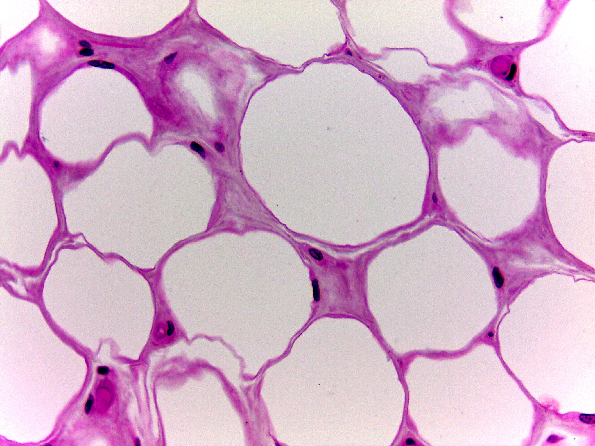
Fat tissue at high power, stained pink and purple. The picture is filled with large, round to many-sided cells that look pale and almost empty, because the fat inside them dissolved when the slide was made. Thin pink walls separate the cells. Where several cells meet, the walls thicken into small patches of pink tissue. Small, dark, flattened or oval nuclei are squeezed against the edges of the cells in these patches. A few small round pink structures with dark crescent-shaped nuclei sit near the corners at the lower left and upper right.